the Creative Commons Attribution 4.0 License.
the Creative Commons Attribution 4.0 License.
 | 27 Jul 2026

A pan-Arctic pigment database for phytoplankton and sea–ice algae

Philipp Assmy
Atreya Basu
Astrid Bracher
Giulia Castellani
Giacomo Ditullio
Katarzyna Dragańska-Deja
Amane Fujiwara
Glaucia Moreira Fragoso
Jacob Høyer
Jiwoon Hwang
Morten Iversen
Anabel von Jackowski
Thomas Juul-Pedersen
Piotr Kowalczuk
Youngju Lee
Maria A. van Leeuwe
Atsushi Matsuoka
Alenya Merz
Christopher John Mundy
Else Ostermann
Ilka Peeken
Matt Pinkerton
Joanna Stoń-Egiert
Jacqueline Stefels
Antonia U. Thielecke
Gaëlle Veyssiere
Hongyan Xi
Eun Jin Yang
Rafael Gonçalves-Araujo
Climate change has dramatically altered the Arctic seas with significant decrease in sea ice extent and thickness and warming water temperature. The ecological impacts of such change have been described for many parts of the Arctic Ocean, but long-term records of biological indicators are still missing. Among those, photosynthetic and accessory pigments are one of the key tools that aid quantification of phytoplankton and sea–ice algae biomass and characterisation of community composition. To address this gap, we present the first pan-Arctic compilation of in situ algal pigment data obtained exclusively by High-Performance Liquid Chromatography (HPLC), containing 10 798 samples collected across 77 Arctic research cruises between 2000 and 2024. As a result of large-scale collaborative effort, this database covers both open water and sea–ice environments across coastal, shelf and open domains. The database (https://doi.org/10.11583/DTU.29445104, Heidemann et al., 2026) includes measures of up to 26 pigments, with 8 major marker/accessory pigments being considered in this study, namely Alloxanthin (Allo), 19'-Butanoyloxyfucoxanthin (But-fuco), Chlorophyll a (Chl a), Chlorophyll b (Chl b), Fucoxanthin (Fuco), 19'-Hexanoyloxyfucoxanthin (Hex-fuco), Peridinin (Peri), and Zeaxanthin (Zea). This publicly available database provides crucial data that can be used to assess phytoplankton dynamics, validating remote sensing observations and can serve as a resource for future Arctic ecological- and modelling studies.
- Article
(4299 KB) - Full-text XML
-
Supplement
(3020 KB) - BibTeX
- EndNote


Climate change has disproportionately affected the Arctic, shown by drastic increases in temperature and ongoing sea–ice decline (England et al., 2021; Stroeve and Notz, 2018). Changing conditions in the Arctic have already altered the food web, with studies indicating further impacts in the near future (Ardyna and Arrigo, 2020; Arrigo and van Dijken, 2015; Flores et al., 2023; Freer et al., 2022; Quinlan et al., 2005). As the foundation of marine food webs, phytoplankton and sea–ice algae are critical for sustaining ecosystems and contribute to large parts of global primary production, leading to energy transfer across trophic levels and ultimately carbon sequestration (Falkowski, 1994; Serra-Pompei et al., 2022). Environmental heterogeneity and changes may alter community composition, with the potential to impact large-scale ecological processes. For example, in the Arctic and sub-Arctic regions, increased open water areas has led to a longer phytoplankton growing season likely associated with changes in community composition. Studies have demonstrated that climate change may favor increased biomass of smaller flagellated phytoplankton species, leading to reduced dominance of large diatoms species (Blais et al., 2017; Coupel et al., 2012; Vonnahme et al., 2025). This can impact the structure of the food web and overall carbon cycling by having an immediate effect on higher trophic levels, potentially influencing the abundance, distribution and feeding preferences of zooplankton (Campbell et al., 2009; Negrete-García et al., 2024).
Characterising phytoplankton community composition is therefore essential for better understanding ecosystem structure, dynamics and services, especially in the under-sampled Arctic seas, which have experienced the greatest impact of climate change in recent years (England et al., 2021). It is clear that long-term data on microalgae physiology and biomass are vital to enhance our ability to assess and detect spatial and temporal patterns in phytoplankton and sea–ice algae community structure and physiology in response to environmental change. Many studies have used pigment-based measurements, but datasets are often scattered across many sources. This makes it difficult to obtain, access and synthesize into a larger spatial and temporal scale. To address this gap, our paper provides baseline information regarding phytoplankton and sea–ice algae biomass and photosynthetic pigments in the Arctic Ocean aiming at providing a framework to support such large-scale Arctic algae studies across two decades of data.
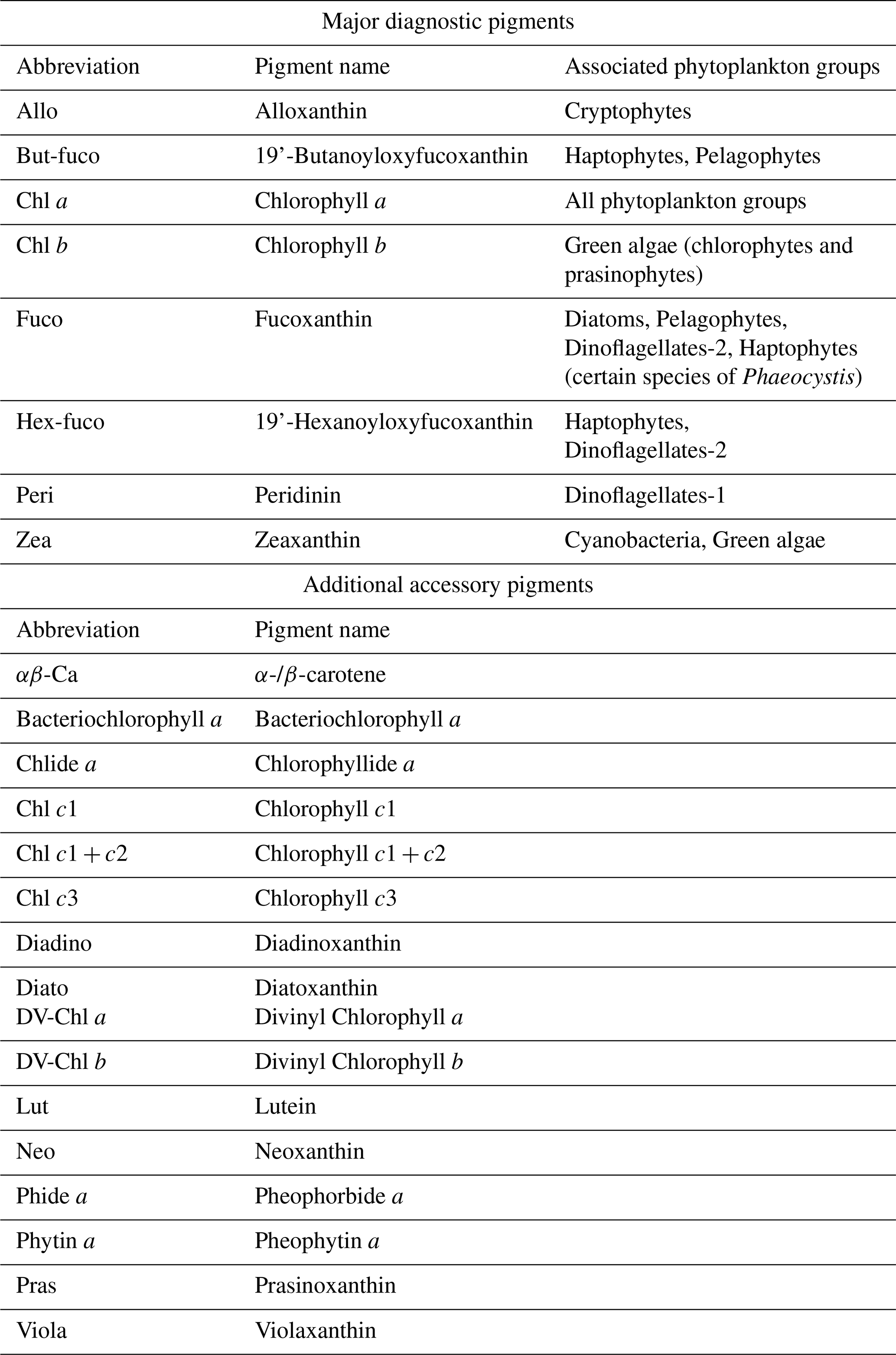
Phytoplankton pigments, particularly chlorophyll a (Chl a), drives photosynthesis by harvesting light and protecting the cells (Falkowski and Kiefer, 1985; Pereira and Gonçalves, 2022). Advances in pigments analysis through High-Performance Liquid Chromatography (HPLC) and ultra HPLC (UHPLC), have enabled precise identification and quantification of a suite of photosynthetic and photoprotective microalgal pigments. These pigments can serve as chemotaxonomic markers for major functional and taxonomic phytoplankton groups, including the commonly used diagnostic pigments, Alloxanthin (Allo), 19'-Butanoyloxyfucoxanthin (But-fuco), Chlorophyll b (Chl b), Fucoxanthin (Fuco), 19'-Hexanoyloxyfucoxanthin (Hex-fuco), Peridinin (Peri), and Zeaxanthin (Zea) used in this study. Additionally and when available, the database presented in this paper also includes α-/β-carotene (αβ-Car), Bacteriochlorophyll a, Chlorophyllide a (Chlide a), Chl c1, Chl c1+c2, Chl-c3, Diadinoxanthin (Diadino), Diatoxanthin (Diato), Divinyl Chlorophyll a and b (DV-Chl a and DV-Chl b), Lutein (Lut), Neoxanthin (Neo), Pheophorbide a (Phide a), Pheophytin a (Phytin a), Prasinoxanthin (Pras) and Violaxanthin (Viola) (Jeffrey et al., 1999) (Table 1). These additional accessory pigments, some of which are also commonly used as marker pigments (Coupel et al., 2015; Fragoso et al., 2017a), enable phytoplankton to adapt and/or acclimate to various light regime changes that occur in the environment (Brunet et al., 2011). Variability in pigment composition can reflect strategies such as photoprotection, which involves the production of pigments that mitigate damage from excessive light and photo acclimation that refers to the ability of algae to adjust their pigment composition in response to light intensity changes that hereby can optimise photosynthetic efficiency (Bonilla et al., 2009; Gosselin et al., 2017).
Table 1Alphabetically ordered list of the major diagnostic pigments and additional accessory pigments considered in this study (including abbreviations, their full name and associated phytoplankton groups) (Fragoso et al., 2017a; Jeffrey et al., 1999). Notice that Dinoflagellates contains two types, reflecting different major pigments.
Absorption and scattering of light by phytoplankton pigments and other cellular constituents influence the reflectance, which allow their detection by radiometers, making them an invaluable source for remote sensing applications (see overview in Bracher et al., 2017). Multispectral satellite sensors such as SeaWiFS, MODIS, MERIS and OLCI have paved the way and demonstrated the ability to distinguish phytoplankton functional types on large scales (Miller, 2004; Nieke et al., 2015). Recent development have focused on hyperspectral sensors, capable of measuring more narrow absorption and reflectance propertied, that can be missed by multispectral sensors (Hu, 2022). Successful applications include ENVISAT's Scanning Imaging Absorption Spectrometer for Atmospheric Chartography (SCIAMACHY) using the PhytoDOAS method (Bracher et al., 2009; Sadeghi et al., 2012) and the DLR Earth Sensing Imaging Spectrometer Mission (DESIS) using the WebAssembly System Interface (WASI) (Gege, 2004, 2014) by Bracher et al. (2021). Most recently NASA's Plankton, Aerosol, Clouds, ocean Ecosystem (PACE) mission have been launched (Werdell et al., 2024) and the European Space Agency (ESA) is preparing the CHIME hyperspectral to be launched in the coming decade (Nieke et al., 2023).
Since satellites provide long-term synoptic observations of phytoplankton groups high-quality in situ pigment data is invaluable for product validation and algorithm refinement (Cetinić et al., 2024). Similarly, several studies based on self-organizing maps, spectral composition or machine-learning-based methods applied to satellite data have leveraged phytoplankton pigments to understand taxonomic shifts in phytoplankton communities (El Hourany et al., 2019, 2024; Hayward et al., 2025; Xi et al., 2020).
While satellite data is becoming increasingly important, it remains limited by its ability to capture only the first optical depth under cloud- and ice-free conditions.
In situ pigment data from the Arctic have compared to other parts of the world historically been limited due to the region's remoteness, harsh weather conditions and logistical challenges of conducting fieldwork in such remote regions. Advances in HPLC technology, together with standardised protocols (Hooker et al., 2005) and round-robins intercomparison of methodologies (Canuti, 2023), have contributed to the growing availability of pigment data globally, which also includes data from the Arctic and Sub-arctic regions (Kramer et al., 2022; Losa et al., 2017; Mattei and Scardi, 2021; Swan et al., 2016; Xi et al., 2023). Related efforts to obtain pigment data, such as those using UPLC methods (Hwang, 2025), also represent a valuable contribution, though they cannot be incorporated into the current database for inter-comparability reasons with HPLC-based measurements. Despite advances, spatial and temporal coverage across the Arctic remains uneven, underscoring persistent knowledge gaps, highlighting the need for coordinated, collective efforts to compile and expand existing data in this rapidly changing region.
To address this gap, we present the first pan-Arctic overview of phytoplankton and sea–ice algae pigment distributions, based on a comprehensive database of Arctic and sub-Arctic in situ pigment measurements collected between 2000 and 2024. The database is compiled from 77 cruises conducted across the Arctic Ocean, covering coastal, shelf, open-ocean and sea–ice domains (see Table 2). Importantly, this database represents a collective effort involving multiple institutions, research teams and international collaborations. By consolidating data from diverse sources, it provides a baseline for assessing long-term trends in phytoplankton pigments, communities and their ecological responses to environmental change. Additionally, such a database can support a pan-Arctic taxonomic characterisation of phytoplankton communities by employing pigment-based taxonomy techniques (Hayward et al., 2023, 2024; Vidussi et al., 2001; Wright and Jeffrey, 2006).
Table 2Alphabetically ordered summary of Arctic and sub-Arctic cruises (=77) with key dataset information such as year, sampling region(s) (as defined in Sect. 3.1.), environment, sample count and associated DOI, where possible. Note that few samples were from melt-ponds, here assigned as ice samples.

2.1 Database compilation
Phytoplankton and ice algae pigment data was compiled from 77 research cruises conducted between 2000 and 2024. The datasets were sourced from a combination of publicly available repositories (e.g., PANGAEA), institutional databases (e.g., NASA) and personal correspondence with scientists. Datasets were accompanied by relevant ancillary metadata, including information on sampling time, location and depth. A total of 10 798 pigment datapoints were compiled (Table 2). Where possible, cruise reports and original publications were consulted to verify the context and methods of data collection. Part of the data has already been published individually for the respective research cruises and/or projects, however, the compiled database presented in this article (including datasets not yet publicly available) has been stored in a public repository (https://doi.org/10.11583/DTU.29445104, Heidemann et al., 2026).
2.2 Data quality measures
This dataset integrated HPLC pigment measurements from 77 research cruises, each carried out by laboratories using their own established laboratory procedures, including different HPLC set-ups, calibration standards and water filtration volume. Methodological descriptions associated with each dataset can be accessed through the provided DOIs in Table 2 or by contacting the original data providers. To ensure consistency and comparability across sources, we applied a set of inclusion/exclusion criteria and harmonisation procedures to ensure data consistency and comparability across sources. Only datasets with measurements of seven out of the eight marker pigments described in Table 1 were included in the database, allowing for reliable inter-sample comparison of pigment composition (criterion 1, see Fig. 1). Although zeaxanthin is normally included in the analysis of phytoplankton functional types, it was excluded as a formal requirement in this database compilation because one source lacked measurement of this pigment and reliable distinction of green algae from other phytoplankton groups remain possible without zeaxanthin (see available pigments for each cruise in Table S1 in the Supplement). Zeaxanthin is also a major diagnostic pigment for Synechococcus, which is largely absent from the Arctic region due to its thermal dependence on warmer waters (Six et al., 2021). Samples were excluded from the final database if they reported Chlorophyll (Chl a) concentration of zero (criterion 2, see Fig. 1), lacked measurements of the required marker pigments and/or were missing essential metadata such as geographic coordinates (criterion 3, see Fig. 1).
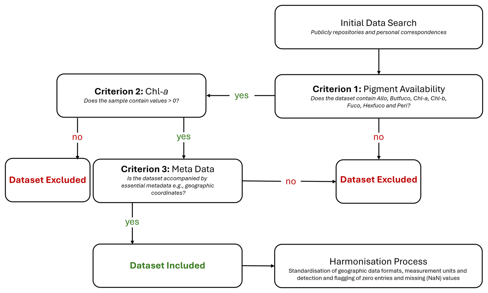
In addition to data filtering, multiple data harmonisation steps were also undertaken: longitudes reported in degrees East (0 to 360° E) were converted to signed longitude (−180 to 180° E/W) to ensure spatial compatibility with standard mapping tools; datasets were also checked for true 0 s and NaNs and that the distinction between these were standardised across datasets. This process required manual inspection of each dataset: If a pigment was entirely absent from a dataset, it was treated as NaNs (empty cells), whereas if a pigment was present but only had a single non-zero value reported, the remaining empty cells were interpreted as true 0s; finally, concentration units for accessory pigments and Chl a were also standardised to the common unit, µg L−1. There was considerable variability in methods used to calculate total Chl a was observed across the database, and these methods were not always reported. Consequently, this parameter was excluded. In addition, only a limited number of melt pond samples were available. These were therefore treated as part of the ice sample category, instead of a category by itself.
2.3 Limitations and uncertainties of the dataset
Despite efforts to minimize both limitations and uncertainties, this dataset consists of HPLC measurements from multiple laboratories. We acknowledge that methodological variability between laboratories exists and will introduce variability to the dataset. The following sections are provided to summarise the main known sources of limitations and uncertainties associated with compiling HPLC data from various laboratories, as well as general uncertainties inherent to pigments measurements.
2.3.1 Analytical
All datasets presented in this paper have followed standard protocols for HPLC analysis of phytoplankton pigments (see Table S2). However, analytical uncertainties can arise from multiple stages of the workflow. As outlined by Wright and Jeffrey (2006), these can broadly be categorised into:
-
Collection and storage: liltration volume, filter type and size, storage temperature and storage time
-
Extraction procedures: sample disruption, duration and temperature, and choice of solvents
-
HPLC methods and techniques(see review of HPLC pigment methods in Roy et al., 2011): variations of column type and flow rate etc.
-
Peak detection, identification, integration and quantification: differences in baseline correction, peak threshold, co-elution handling and pigment standards used for calibration
To minimize bias in each of these steps, substantial effort has been made through inter-comparability studies. Most notably are the SeaWiFS HPLC Analysis Round-Robin Experiments (SeaHarre-1 to SeaHarre-5) (Hooker et al., 2012) and HPLC/DAD intercomparison on phytoplankton pigments (HIP-1 to HIP-4) (Artuso et al., 2016; Canuti et al., 2016), which efforts has been to eliminate differences in pigment quantification across laboratories and quantify these uncertainties in terms of what can be expected, even when conducting analysis according to strict protocols.
2.3.2 Pigment-specific
In addition to the analytical sources of uncertainties, pigment-specific sources also exist because of a varying degree of sensitivity to the above-described workflow. Any chosen HPLC method can therefore increase or reduce the variance experienced for a specific pigment. However, defining robust “uncertainty values” for each individual pigment remains a challenge across analytical methods. Recent inter-laboratory comparisons highlight this challenge, while also underscoring the need of such estimates in applications such as remote sensing validation (e.g., Canuti, 2023).
A main source of pigment-specific behavior comes from differences in the chemical stability of the pigments. This will affect their susceptibility to degradation, co-elution and extraction efficiency. For example, Chlorophylls (a, b, c1 and c2) are prone to degradation, particularly during storage and extraction, producing degradation products such as pheophytins and chlorophyllides. This can lead to biased and unprecise chlorophyll estimations as well as co-elution issues. This have often been reported for chlorophyll c1 and chlorophyll c2 as well as for the Fuco derivatives (But-fuco and Hex-fuco) (Roy et al., 2011; Simmons et al., 2016; Wright and Jeffrey, 2006; Zapata et al., 2000). Additionally, some chlorophyll degradation products, for example Phide a and Phytin a, can co-elute with carotenoids due to polarity changes (Mendes et al., 2007).
Finally, pigment concentration itself can be a major source of uncertainty. This is particularly true for low-abundance accessory pigments, which often approach the instrumental limits of detection (LOD) (Bidigare et al., 2002; Canuti, 2023; Canuti et al., 2016; Claustre et al., 2004; Hooker et al., 2005). For instance, when pigment concentrations fall below 0.01 mg m−3, the relative standard deviation can reach up to nearly double that of pigments present at higher concentrations (Claustre et al., 2004).
3.1 Sampling coverage
To classify the sampling efforts across the Arctic and sub-Arctic seas into common groups, the study area was subdivided into 15 regions (Fig. 2). A total of 10 095 water samples and 703 sea–ice samples were compiled and mapped according to their geographic location. Water samples displayed extensive sampling across most regions, while ice samples were comparatively sparse and primarily concentrated in the Central Arctic Ocean (CAO), Canadian Archipelago and Hudson Bay. These samples originate from a limited number of cruises and are not consistently represented throughout the years. Regional sampling frequencies are summarised in Fig. 2, highlighting the dominance of water samples and the uneven spatial distribution of sea–ice samples.
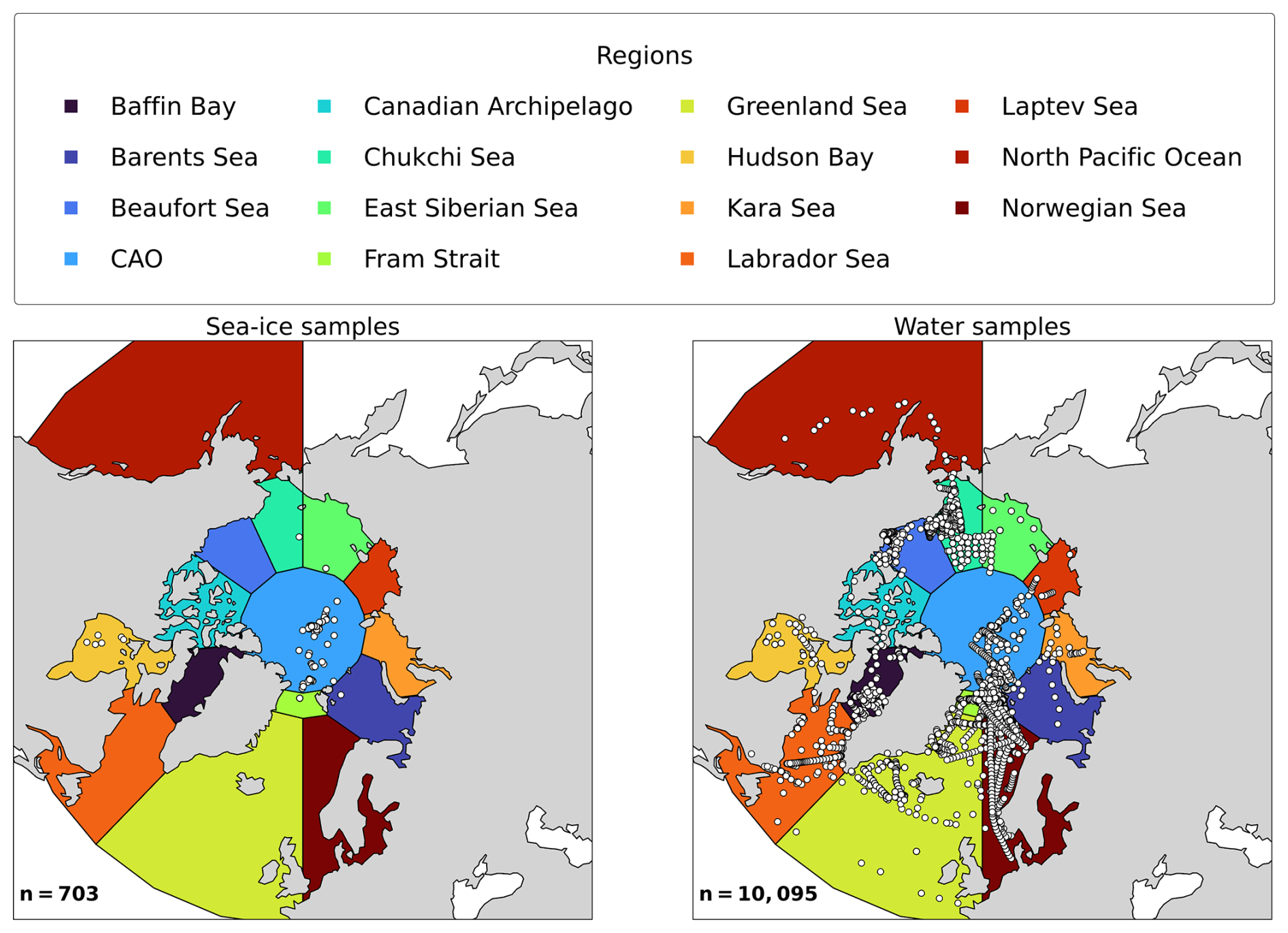
Figure 2Map showing the spatial distribution of water and sea–ice samples across 15 defined regions of the Arctic and sub-Arctic seas. Each point represents an individual sampling location.
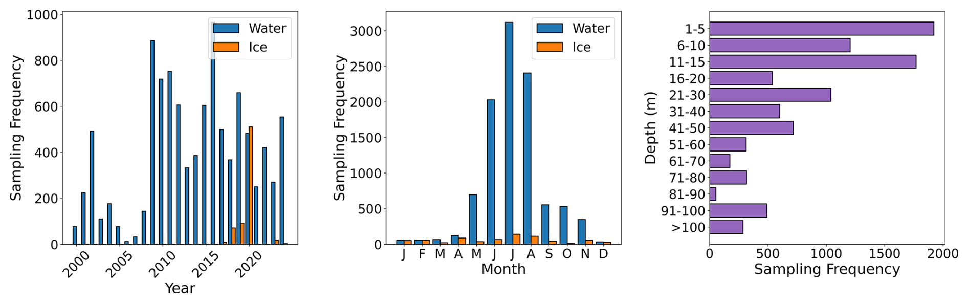
Figure 3Leftpanel: yearly sampling frequency for water column and sea–ice samples, middle panel: monthly sampling frequency and right panel: number of water samples observations per depth bin. Note that for sea–ice samples, the vertical distribution is not presented due to inconsistencies in the ice core sampling methods among the different campaigns.
Overall, the water sample collection also varied substantially across years, with higher coverage in 2002, 2009, 2016 and 2020 (Fig. 3). These periods correspond to large-scale, multidisciplinary campaigns such as MALINA (Coupel et al., 2015), GreenEdge (Bruyant et al., 2022a; Massicotte et al., 2020), the AR7W transect during the MOSAiC cruise and others, including global initiatives such as the International Polar Year (IPY). Additionally, the MAREDAT pigment database represents a major synthesis effort (Peloquin et al., 2013a) (see Table 1).
Seasonally, sampling frequency was the highest in spring and summer (May–August). Sampling frequency was lower in September, followed by a notable secondary drop in December. The lowest sampling frequency is observed during winter and early spring (December–March), when there is extensive sea ice extent. Sampling frequency also varied vertically in the water column (Fig. 3). Most samples were collected within the upper 1–15 m range. Sampling frequency consistently declines from 21–30 m onward, before a slight increase in the 91–100 m range.
3.2 Pigment distribution
Pigment concentration in water and sea–ice samples exhibited considerable variability in the biomass and composition of algal communities as well as in the spatiotemporal structure of the database (Table 3). In addition, the observed variability can also be attributed to adaptive pigment strategies.
Table 3Summary of 5th, 95th, mean, median and std statistics across all cruises (=77), for (a) pigment concentration categorized by sample type and (b) relative ratio of accessory pigments vs. Chl a, For detailed pigment information by individual cruises, see Table S1. For detailed information about all pigments across environments, see Table A1.
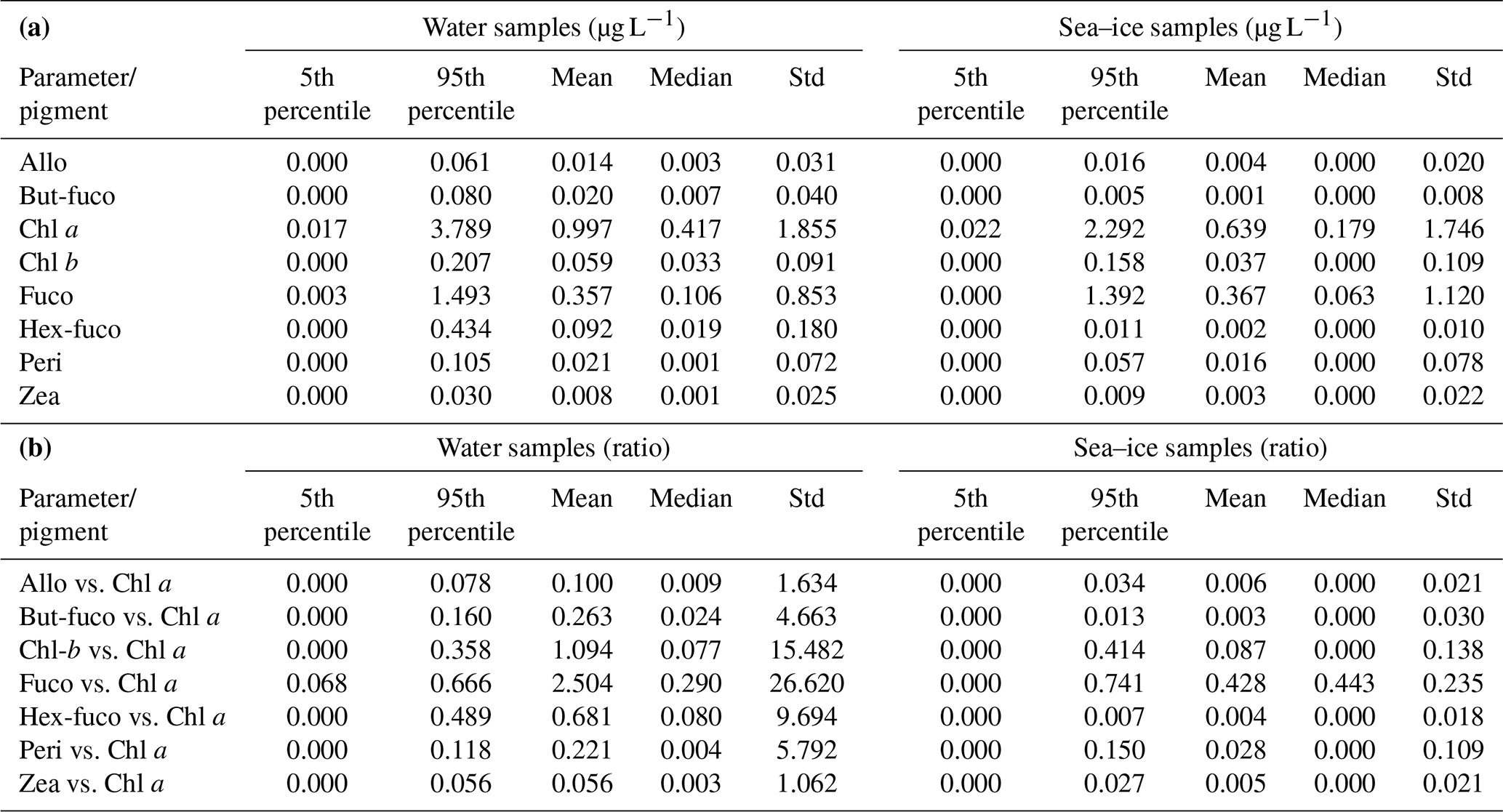
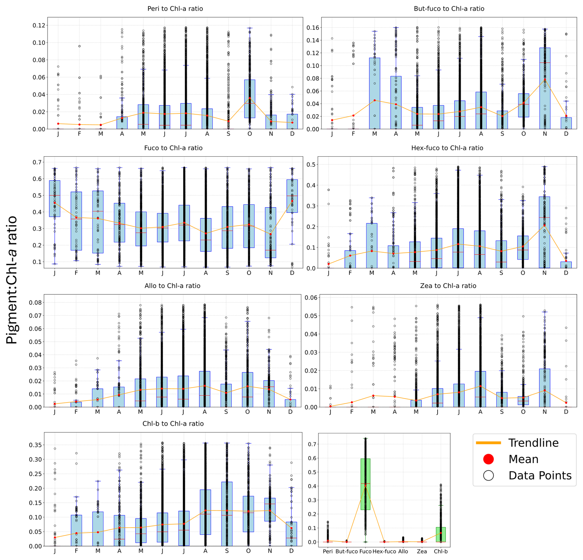
Figure 4Display of the seasonal variation of individual accessory pigment concentration to Chl a ratio. Water samples are shown as monthly ratios in blue and sea–ice samples are shown overall across months per pigment in green. Only values within 5th to 95th percentile are included. Data points are displayed as open circles, the mean ratio for each month is shown as a red dot and an orange line connects means.
Since direct comparison of absolute pigments concentrations between sea–ice and water samples should be interpreted with caution, only a brief description of absolute pigment concentration is provided for context (Table 3a). Chl a, the primary indicator of phytoplankton and sea–ice algae biomass, had a mean concentration of 0.997 µg L−1 in water samples and 0.639 µg L−1 in sea–ice samples although the highest individual values in both environments point to episodic blooms. These values represent the mean of all individual concentration measurements in the compiled dataset. The highest concentration across the database, 32.76 µg L−1, was recorded in the Chukchi Sea originating from the Ice Scape dataset in August 2010. In general, such high concentrations are rare, with the majority of samples falling below 0.5 µg L−1 across seasons. Overall, concentrations were highly variable with 90 % of values falling between 0.017 and 3.789 µg L−1 in water samples and between 0.022 and 0.292 µg L−1 in sea–ice samples (Table 3a). It should be recognised that there is a potential bias when comparing open-water and bulk–ice samples due to neglect of the dilution of pigments when bulk sea–ice cores are melted, which can underestimate concentrations from sympagic algae (Chamberlain et al., 2022; Gradinger, 2009; Miller et al., 2015).
In terms of the relative ratios to Chl a (Table 3b and Fig. 4) the database also showed marked seasonal variability. Among the eight analysed marker pigments, Fuco consistently had the highest ratios, particularly during winter months, followed by a decline through summer and early autumn. The Fuco-derivatives, But-fuco and Hex-fuco, both considered diagnostic markers of haptophytes, had a stable distribution throughout the year, aside from notable peaks in November.
The ratio of Allo, a distinct marker pigment of cryptophytes, started to increase in late winter, prior to the main growing season. Peri, which is a marker pigment of dinoflagellates, had low overall ratios. These ratios increased during summer, peaking in October, before they exhibited a sharp decline toward the onset of December. As expected, Zea and Chl b were relatively aligned throughout most of the year, consistent with their shared role as a marker pigment of green algae. However, a marked dissimilarity in their patterns occurred in November, when Chl b declined while Zea increased.
For the sea–ice samples, the only accessory pigments with high ratios are Fuco and Chl b.
These differences suggest distinct pigment signatures between sea–ice and water samples, underscoring the importance of expanding sea–ice sampling efforts to better capture algae dynamics in this environment.
3.2.1 Spatial and temporal dynamics
In our dataset, Chl a exhibited strong spatial and temporal (seasonal) and inter-annual variability, reflecting the dynamic nature of phytoplankton and sea–ice algae in polar and subpolar environments. Elevated concentrations were observed in areas such as the Fram Strait, Barents Sea, Beaufort Sea, Chukchi Sea and Labrador Sea (Fig. 5). However, as the database is opportunistic by nature, covering only discrete regional samplings, spatial and seasonal comparisons should be made with caution.
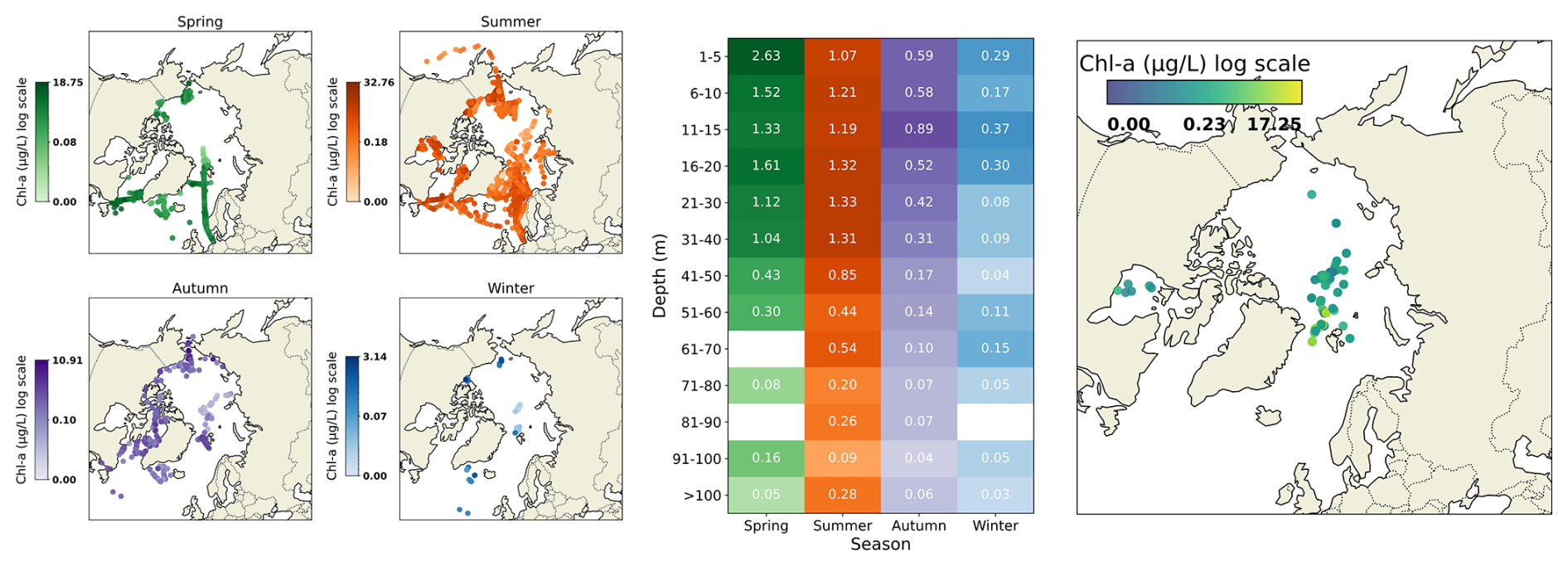
Figure 5(a) Spatial distribution of all measured Chl a water samples in each season, shown as subplots for spring, summer, autumn and winter. Color mapping uses logarithmic scale, but the values themselves are not log-transformed. (b) Mean Chl a concentration as function of depth range (rows) and season (columns). Color indicates the average value for each depth/season bin. Missing color indicates that data is not available. (c) Spatial distribution of all measured Chl a for sea–ice samples.
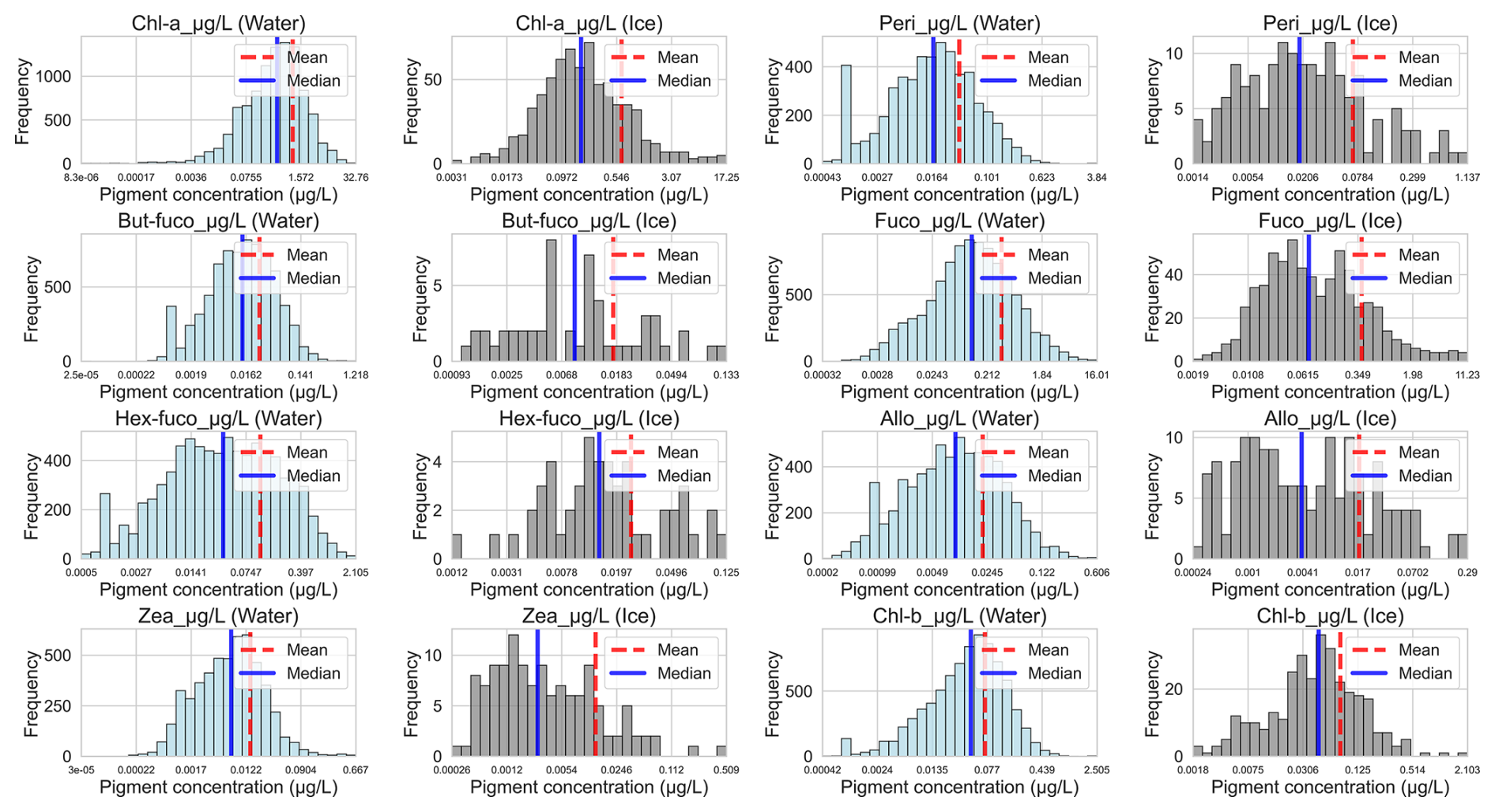
Figure 6Frequency histograms displaying log-scaled pigment concentrations. Water samples are shown in blue and sea–ice samples are shown in grey. x axis represents actual pigment concentrations. Note the different ranges for y axis comparing water and sea–ice samples.
In spring, the Labrador Sea recorded the seasonal maximum (18.75 µg L−1), with the highest values occurring mostly in surface waters (0–5 m). As previously mentioned, summer exhibited the maximum database record for Chl a. Contrary to spring, highest averages were found between 16–30 m. By autumn, Chl a had declined across most regions. However, localised high biomass persisted in some areas such as Beaufort Sea, Chukchi Sea and Fram Strait. Winter Chl a was generally low.
The concentration frequency distribution plots of water samples (Fig. 6, blue plots) showed that both Chl a, Chl b and Fuco exhibited approximately log-normal distribution, with peak concentrations near central values and tails on either side. This interpretation was supported by the log-normal distribution of data. However, formal normality tests such as Kolmogorov–Smirnov (K–S test) indicated significant deviations from perfect log-normality, likely due to large sample size and/or the presence of outliers (see Fig. A1). In contrast, pigments such as Zea, Peri and the Fuco derivatives exhibited more variable distributions and greater heterogeneity among samples. The histograms for the sea–ice samples (Fig. 6, grey plots), had much more variability in the distribution of concentrations. This is likely also related to smaller sample size for sea–ice samples.
All datasets used in this paper have been compiled into a single repository, accessible using https://doi.org/10.11583/DTU.29445104 (Heidemann et al., 2026).
Photosynthetic and accessory pigments serve as indicators of community structure and biomass, and are important oceanographic variables for understanding ocean ecology, as they can also support assessment of biogeochemical cycling. Yet, datasets reporting concentrations of those pigments remain sparse in the Arctic region. To address this gap, we compiled, filtered, standardised and harmonised 10 798 pigment samples from HPLC, including water (n=10 095) and sea–ice samples (n=703), collected during 77 research cruises spanning from 2000 to 2024 across 15 defined Arctic and sub-Arctic regions. Sampling frequency varied by year, with peak efforts in 2002, 2009, 2016 and 2020. These years correspond to major multidisciplinary research projects and international initiatives such as the International Polar Year (IPY). Seasonally, most samples were collected during spring and summer months in the upper water column.
Within the available dataset, Chl a was found in higher concentrations in sea–ice samples compared to water samples, on average. However, for accessory pigments, concentrations were generally found to be lower in sea–ice samples. Despite this, the highest concentration of Chl a across the dataset, 32.76 µg L−1, was recorded for a water sample in the Chukchi Sea. Other areas that exhibited elevated Chl a concentrations were spread across the Arctic marginal seas such as the Fram Strait, Barents Sea, Beaufort Sea and Labrador Sea. Interestingly, among the eight marker pigments, Fuco consistently had the highest pigment to Chl a ratios, particularly during winter months, followed by a decline through summer and early autumn. For the sea–ice samples, the only accessory pigments with high ratios were Fuco and Chl b.
Our comprehensive phytoplankton and sea–ice algal HPLC-based pigment dataset provides valuable insights into the research effort in the Arctic Ocean. Furthermore, it offers important observations on spatial and temporal patterns of primary producers supporting large-scale environmental research in the Arctic across over two decades of sampling efforts.
Table A1Pigment concentration statistics for all included measured pigments, separated by sample type and summarised across all cruises. For each pigment and environment, the table reports the 5th percentile, 95th percentile, mean, median and standard deviation (Std).
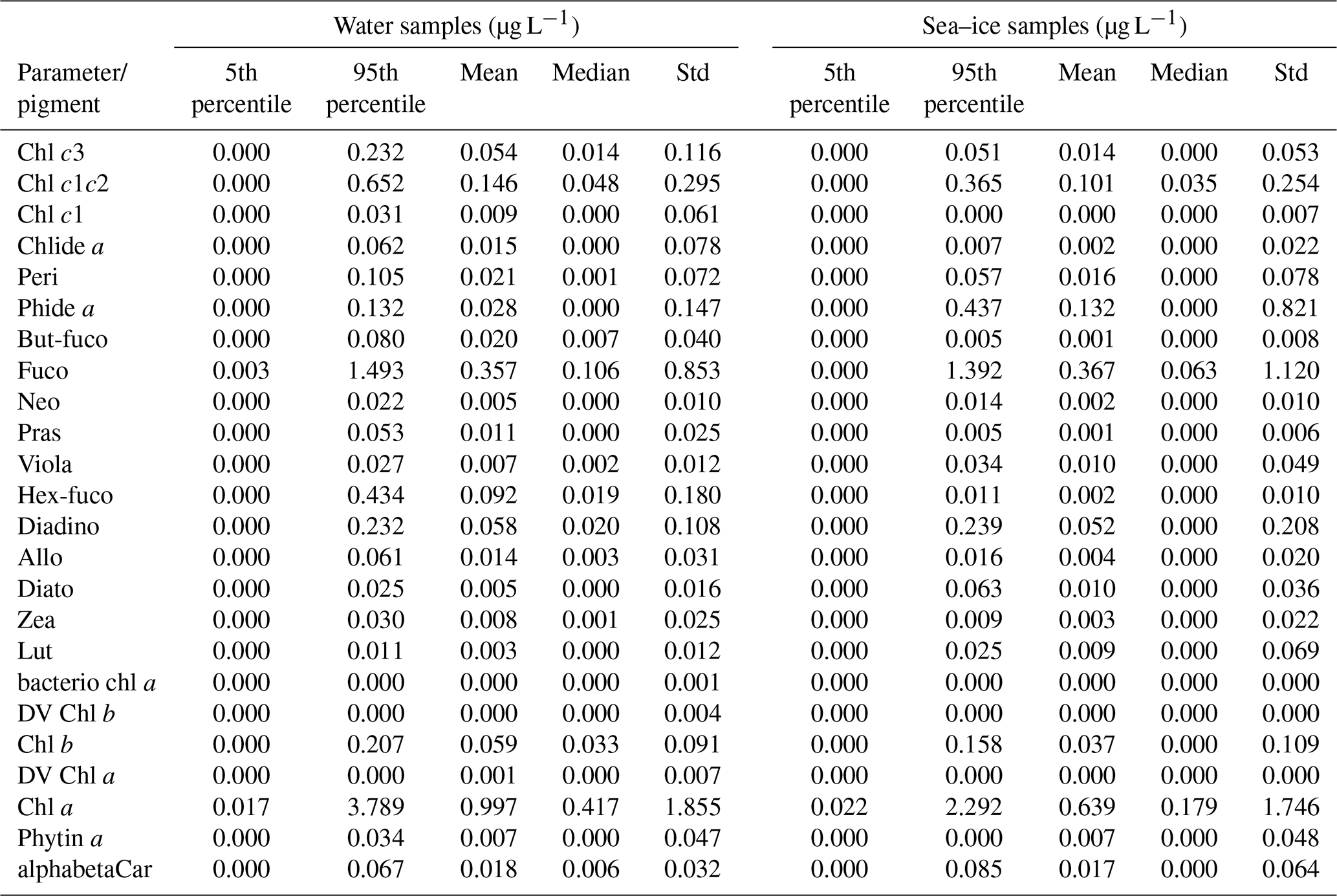
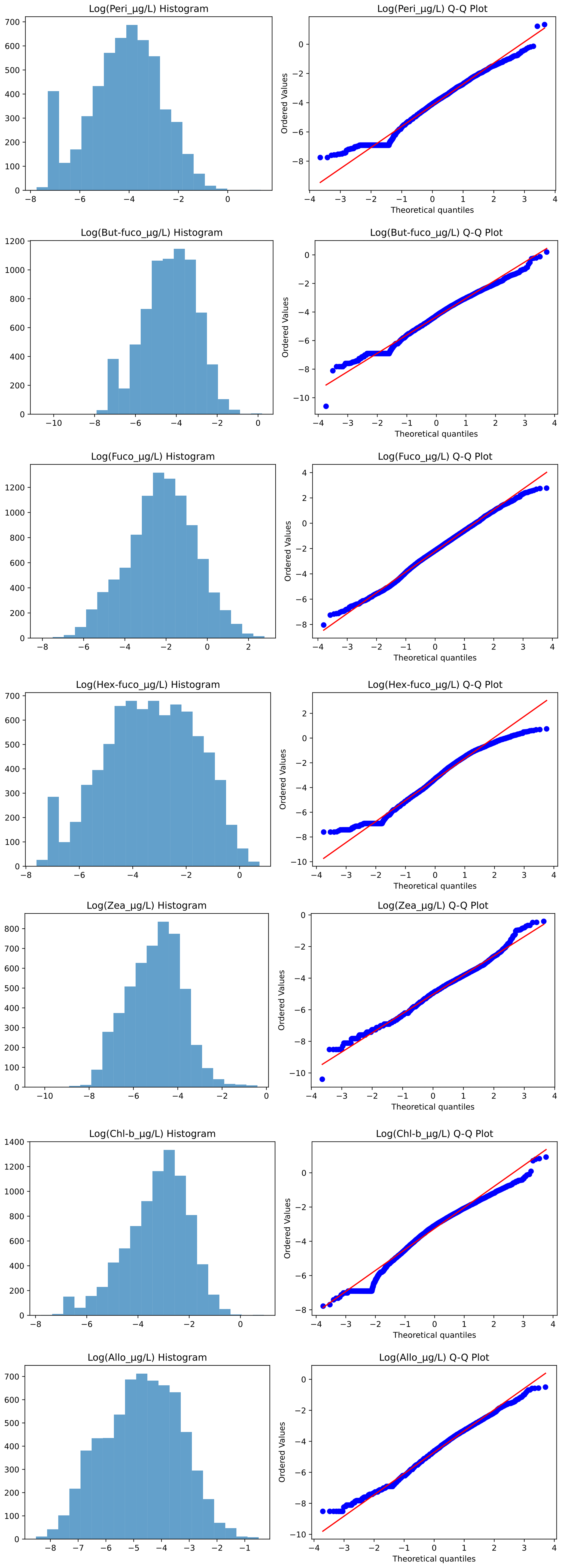
Figure A1Histogram and Q–Q plots performed for each marker pigment on standardised and log-transformed data. For each pigment, a histogram and a Q–Q plot are visualized to assess normality. All Kolmogorov–Smirnov (K–S) p values were below 0.05 test, indicating that the data deviates from a normal distribution.
The supplement related to this article is available online at https://doi.org/10.5194/essd-18-5505-2026-supplement.
ACH consolidated the data, harmonized and standardised the dataset, performed the data analysis, generated plots and developed the manuscript. AH conceived the study idea, provided relevant expertise, contributed data, facilitated key contacts and data sources and contributed to revising and improving the data analysis and manuscript. RGA provided relevant expertise and contributed to conceiving the study idea, revising and improving the data analysis and manuscript. PA, AsB, GC, GD, KDD, AF, GMF, JaH, MI, AJ, TJP, PK, YL, MAVL, AtM, AlM, CJM, EO, IP, MP, JSE, JS, AT, GV, HX and EJY contributed data, provided relevant expertise and revised the manuscript. AtB and JiH provided relevant expertise and revised the manuscript.
The contact author has declared that none of the authors has any competing interests.
Publisher's note: Copernicus Publications remains neutral with regard to jurisdictional claims made in the text, published maps, institutional affiliations, or any other geographical representation in this paper. The authors bear the ultimate responsibility for providing appropriate place names. Views expressed in the text are those of the authors and do not necessarily reflect the views of the publisher.
We thank the captain, the crew and all scientists of R/Vs Maria S. Merian MSM93 and Polarstern expeditions PS93-2, PS99-2, PS107, PS121, PS126, PS131, PS136 and PS143-2 for their support in preparation and on board the expeditions.
We gratefully acknowledge the researchers, institutions, and scientific programs that have made their datasets publicly available through open-access repositories, as well as our collaborators and co-authors who contributed additional datasets. This study would not have been possible without the collective commitment of the scientific community to openly share and preserve high-quality observations. Such efforts are fundamental to advancing collaboration, reproducibility, and scientific discovery, and we hope this work highlights the lasting value of open data for the broader research community.
Asta C. Heidemann and Rafael Gonçalves-Araujo were supported by the Independent Research Fund Denmark, through the project WaterColor (grant no. 4251-00058B). Rafael Gonçalves-Araujo was also supported by the European Union's Horizon Europe research and innovation program under Grant Agreement no. 101136480 (SEA-Quester).
Philipp Assmy was supported by the Research Council of Norway (project no. 244646) and the Ministry of Foreign Affairs, Norway, through the ID Arctic project. Ship time onboard R/V Lance during the N-ICE2015 expedition was provided by the Norwegian Polar Institute.
Atsushi Matsuoka was supported by NASA PACE (grant no. 80NSSC26K0091) and JAXA's GCOM-C/SGLI (25RT000334) projects.
Youngju Lee was supported by Korea Institute of Marine Science and Technology Promotion (KIMST) grant funded by the Ministry of Oceans and Fisheries (KIMST RS-2021-KS211500, Korea-Arctic Ocean Warming and Response of Ecosystem, KOPRI).
Katarzyna Dragańska-Deja, Joanna Stoń-Egiert and Piotr Kowalczuk were supported by a project funded by the Polish National Science Centre (NCN) OPUS26 project OptiCal-Green (2023/51/B/ST10/01344) granted to Piotr Kowalczuk. Additional funding has been provided by the European Union's Horizon Europe research and innovation program under Grant Agreement no. 101136480 (SEA-Quester) and under Grant Agreement no. 101136748 (BioEcoOcean), as well as the statutory research program of the Institute of Oceanology, Polish Academy of Sciences (tasks I.2 and II.5). The ship time on board of r/v Kronprins Haakon was provided by the Norwegian Polar Institute, Tromsø, Norway.
Glaucia Moreira Fragoso was supported by Fisheries and Oceans Canada (DFO). We acknowledge the Atlantic Zone Offshelf Monitoring Program, the captains and crew of the Canadian Coast Guard vessel CCGS Hudson, and DFO staff for their contributions to data collection during the Labrador Sea cruises and laboratory analysis at the Bedford Institute of Oceanography.
Astrid Bracher and Hongyan Xi were supported by the Helmholtz Infrastructure Initiative FRAM, the Deutsche Forschungsgemeinschaft (DFG, German Research Foundation) – Projektnummer 268020496 – TRR 172, within the “Transregional Collaborative Research Center ArctiC Amplification: Climate Relevant Atmospheric and SurfaCe Processes and Feedback Mechanisms (AC)3” (Project C03), the Copernicus Marine – Innovation Service Evolution R&D Project ML-PhyTAO (23138L03D-COP-INNO SCI-9000), and the ESA project Phyto-CCI (4000147645/25/I-LR). Alexander Hayward was also supported by the ESA project Phyto-CCI (4000147645/25/I-LR). Ship time for Astrid Bracher's and Hongyna Xi's pigment data sampling was provided under grant numbers AWI_PS93.2_02, AWI_PS99_00, AWI_PS107_09, AWI_PS121_02, AWI_PS126_02, AWI_PS131_05, AWI_PS136_04, AWI_PS143.2_02 and GPF 18-1_33.
Ilka Peeken was supported by the PoF III & IV program providing ship time on R/V Polarstern [Changing Earth – Sustaining our Future, Topic 6.1 of the Helmholtz Association].
Antonia U. Thielecke was supported by the Helmholtz Young Investigator Group SiDe-Effect (VH-NG-1600) awarded to Mar Fernández-Méndez. PS138 (ArcWatch I) was supported by the Helmholtz Research Programme “Changing Earth – Sustaining our Future” Topic 6, Subtopics 1, 2 and 3 (grant no. AWI_PS138_02).
Jacqueline Stefels and Maria A. van Leeuwe were supported by the Dutch Research Council (NOW), through the Netherlands Polar Programme (NPP), Project no 866.18.002.
This paper was edited by Alexander Fraser and reviewed by Vanda Brotas and one anonymous referee.
Ardyna, M. and Arrigo, K. R.: Phytoplankton dynamics in a changing Arctic Ocean, Nat. Clim. Change, 10, 892–903, https://doi.org/10.1038/s41558-020-0905-y, 2020.
Arrigo, K. R. and van Dijken, G. L.: Continued increases in Arctic Ocean primary production, Synth. Arct. Res. SOAR, 136, 60–70, https://doi.org/10.1016/j.pocean.2015.05.002, 2015.
Artuso, F., Canuti, E., Cataldi, D., Costa Goela, P., Grung, M., Ras, J., and Röttgers, R.: HPLC/DAD intercomparison on phytoplankton pigments (HIP-1, HIP-2, HIP-3 and HIP-4), Publications Office of the European Union, https://doi.org/10.2788/134904, 2016.
Assmy, P., Duarte, P., Dujardin, J., Fernández-Méndez, M., Fransson, A., Hodgson, R., Kauko, H., Kristiansen, S., Mundy, C., Olsen, L. M., Peeken, I., Sandbu, M., Wallenschus, J., and Wold, A.: N-ICE2015 water column biogeochemistry, Norwegian Polar Data Centre [data set], https://doi.org/10.21334/NPOLAR.2016.3EBB7F64, 2016.
Bidigare, R. R., Van Heukelem, L., and Trees, C. C.: HPLC phytoplankton pigments: sampling, laboratory methods, and quality assurance procedures, Ocean Opt. Protoc. Satell. Ocean Color Sens. Valid. Revis., 3, 258–268, 2002.
Blais, M., Ardyna, M., Gosselin, M., Dumont, D., Bélanger, S., Tremblay, J.-É., Gratton, Y., Marchese, C., and Poulin, M.: Contrasting interannual changes in phytoplankton productivity and community structure in the coastal Canadian Arctic Ocean, Limnol. Oceanogr., 62, 2480–2497, https://doi.org/10.1002/lno.10581, 2017.
Bonilla, S., Rautio, M., and Vincent, W. F.: Phytoplankton and phytobenthos pigment strategies: implications for algal survival in the changing Arctic, Polar Biol., 32, 1293–1303, https://doi.org/10.1007/s00300-009-0626-1, 2009.
Bracher, A.: Phytoplankton pigment concentration and phytoplankton groups measured on water samples obtained during POLARSTERN cruise PS106 in the Arctic Ocean, Alfred Wegener Institute, Helmholtz Centre for Polar and Marine Research, Bremerhaven, PANGAEA [data set], https://doi.org/10.1594/PANGAEA.899284, 2019.
Bracher, A. and Wiegmann, S.: Phytoplankton pigment concentrations during POLARSTERN cruise PS121 from North Sea to Fram in August to September 2019, PANGAEA [data set], https://doi.org/10.1594/PANGAEA.941011, 2022.
Bracher, A., Vountas, M., Dinter, T., Burrows, J. P., Röttgers, R., and Peeken, I.: Quantitative observation of cyanobacteria and diatoms from space using PhytoDOAS on SCIAMACHY data, Biogeosciences, 6, 751–764, https://doi.org/10.5194/bg-6-751-2009, 2009.
Bracher, A., Bouman, H. A., Brewin, R. J. W., Bricaud, A., Brotas, V., Ciotti, A. M., Clementson, L., Devred, E., Di Cicco, A., Dutkiewicz, S., Hardman-Mountford, N. J., Hickman, A. E., Hieronymi, M., Hirata, T., Losa, S. N., Mouw, C. B., Organelli, E., Raitsos, D. E., Uitz, J., Vogt, M., and Wolanin, A.: Obtaining Phytoplankton Diversity from Ocean Color: A Scientific Roadmap for Future Development, Front. Mar. Sci., 4, https://doi.org/10.3389/fmars.2017.00055, 2017.
Bracher, A., Soppa, M. A., Gege, P., Losa, S. N., Silva, B., Steinmetz, F., and Droscher, I.: Extension of Atmospheric Correction Polymer to Hyperspectral Sensors: Application to HICO and First Results for DESIS Data, in: 2021 IEEE International Geoscience and Remote Sensing Symposium IGARSS, https://doi.org/10.1109/IGARSS47720.2021.9553568, 2021.
Bracher, A., Xi, Ho., Hohe, C., Wiegmann, S., and Peeken, I.: Phytoplankton pigment and phytoplankton group chlorophyll-a concentrations during POLARSTERN cruise PS126 from North Sea to Fram Strait in May to June 2021 from HPLC analysis of water samples, PANGAEA [data set], https://doi.org/10.1594/PANGAEA.982954, 2025a.
Bracher, A., Xi, H., and Hohe, C.: Phytoplankton pigment and phytoplankton group chlorophyll-a concentrations during POLARSTERN cruise PS131 from North Sea to East Greenland Sea in 29. June to 3. August 2022 from HPLC analysis of water, PANGAEA [data set], https://doi.org/10.1594/PANGAEA.983242, 2025b.
Bracher, A., Xi, H., and Wiegmann, S.: Phytoplankton pigment and phytoplankton group chlorophyll-a concentrations during POLARSTERN cruise PS136 from North Sea to Fram Strait in May to June 2023 from HPLC analysis of water samples, PANGAEA [data set], https://doi.org/10.1594/PANGAEA.983243, 2025c.
Bracher, A., Xi, H., and Wiegmann, S.: Phytoplankton pigment and phytoplankton group chlorophyll-a concentrations during POLARSTERN cruise PS143/2 from Tromsø to Fram Strait in 12 July to 5 August 2024 from HPLC analysis of water samples, PANGAEA [data set], https://doi.org/10.1594/PANGAEA.983244, 2025d.
Bracher, A., Hohe, C., Wiegmann, S., and Xi, H.: Phytoplankton pigment concentrations during Maria S. Merian cruise MSM93 from North Sea to Fram Strait and back in June to July 2020 from HPLC analysed water samples, PANGAEA [data set], https://doi.org/10.1594/PANGAEA.995973, 2026.
Brunet, C., Johnsen, G., Lavaud, J., and Roy, S.: Pigments and photoacclimation processes, in: Phytoplankton Pigments: Characterization, Chemotaxonomy and Applications in Oceanography, https://hal.science/hal-01101814 (last access: 9 July 2026), 2011.
Bruyant, F., Amiraux, R., Amyot, M.-P., Archambault, P., Artigue, L., Barbedo de Freitas, L., Bécu, G., Bélanger, S., Bourgain, P., Bricaud, A., Brouard, E., Brunet, C., Burgers, T., Caleb, D., Chalut, K., Claustre, H., Cornet-Barthaux, V., Coupel, P., Cusa, M., Cusset, F., Dadaglio, L., Davelaar, M., Deslongchamps, G., Dimier, C., Dinasquet, J., Dumont, D., Else, B., Eulaers, I., Ferland, J., Filteau, G., Forget, M.-H., Fort, J., Fortier, L., Galí, M., Gallinari, M., Garbus, S.-E., Garcia, N., Gérikas Ribeiro, C., Gombault, C., Gourvil, P., Goyens, C., Grant, C., Grondin, P.-L., Guillot, P., Hillion, S., Hussherr, R., Joux, F., Joy-Warren, H., Joyal, G., Kieber, D., Lafond, A., Lagunas, J., Lajeunesse, P., Lalande, C., Larivière, J., Le Gall, F., Leblanc, K., Leblanc, M., Legras, J., Lévesque, K., Lewis, K.-M., Leymarie, E., Leynaert, A., Linkowski, T., Lizotte, M., Lopes dos Santos, A., Marec, C., Marie, D., Massé, G., Massicotte, P., Matsuoka, A., Miller, L. A., Mirshak, S., Morata, N., Moriceau, B., Morin, P.-I., Morisset, S., Mosbech, A., Mucci, A., Nadaï, G., Nozais, C., Obernosterer, I., Paire, T., Panagiotopoulos, C., Parenteau, M., Pelletier, N., Picheral, M., Quéguiner, B., Raimbault, P., Ras, J., Rehm, E., Ribot Lacosta, L., Rontani, J.-F., Saint-Béat, B., Sansoulet, J., Sardet, N., Schmechtig, C., Sciandra, A., Sempéré, R., Sévigny, C., Toullec, J., Tragin, M., Tremblay, J.-É., Trottier, A.-P., Vaulot, D., Vladoiu, A., Xue, L., Yunda-Guarin, G., and Babin, M.: The Green Edge cruise: investigating the marginal ice zone processes during late spring and early summer to understand the fate of the Arctic phytoplankton bloom, Earth Syst. Sci. Data, 14, 4607–4642, https://doi.org/10.5194/essd-14-4607-2022, 2022a.
Bruyant, F., Amiraux, R., Amyot, M.-P., et al.: The Green Edge cruise: following the evolution of the Arctic phytoplankton spring bloom, from ice-covered to open waters, SEANOE [data set], https://doi.org/10.17882/86417, 2022b.
Campbell, R. G., Sherr, E. B., Ashjian, C. J., Plourde, S., Sherr, B. F., Hill, V., and Stockwell, D. A.: Mesozooplankton prey preference and grazing impact in the western Arctic Ocean, Deep-Sea Res. Pt. II, 56, 1274–1289, https://doi.org/10.1016/j.dsr2.2008.10.027, 2009.
Canuti, E.: Phytoplankton pigment in situ measurements uncertainty evaluation: an HPLC interlaboratory comparison with a European-scale dataset, Front. Mar. Sci., 10, https://doi.org/10.3389/fmars.2023.1197311, 2023.
Canuti, E., Grung, J., Röttgers, M., Goela, C., and Artuso, P.: HPLC/DAD Intercomparison on Phytoplankton Pigments (HIP-1, HIP-2, HIP-3 and HIP-4), European Union, https://doi.org/10.2788/47099, 2016.
Cetinić, I., Rousseaux, C. S., Carroll, I. T., Chase, A. P., Kramer, S. J., Werdell, P. J., Siegel, D. A., Dierssen, H. M., Catlett, D., Neeley, A., Soto Ramos, I. M., Wolny, J. L., Sadoff, N., Urquhart, E., Westberry, T. K., Stramski, D., Pahlevan, N., Seegers, B. N., Sirk, E., Lange, P. K., Vandermeulen, R. A., Graff, J. R., Allen, J. G., Gaube, P., McKinna, L. I. W., McKibben, S. M., Binding, C. E., Calzado, V. S., and Sayers, M.: Phytoplankton composition from sPACE: Requirements, opportunities, and challenges, Remote Sens. Environ., 302, 113964, https://doi.org/10.1016/j.rse.2023.113964, 2024.
Chamberlain, E. J., Balmonte, J. P., Torstensson, A., Fong, A. A., Snoeijs-Leijonmalm, P., and Bowman, J. S.: Impacts of sea ice melting procedures on measurements of microbial community structure, Elem. Sci. Anthr., 10, 00017, https://doi.org/10.1525/elementa.2022.00017, 2022.
Claustre, H., Hooker, S. B., Van Heukelem, L., Berthon, J.-F., Barlow, R., Ras, J., Sessions, H., Targa, C., Thomas, C. S., van der Linde, D., and Marty, J.-C.: An intercomparison of HPLC phytoplankton pigment methods using in situ samples: application to remote sensing and database activities, Mar. Chem., 85, 41–61, https://doi.org/10.1016/j.marchem.2003.09.002, 2004.
Coupel, P., Jin, H. Y., Joo, M., Horner, R., Bouvet, H. A., Sicre, M.-A., Gascard, J.-C., Chen, J. F., Garçon, V., and Ruiz-Pino, D.: Phytoplankton distribution in unusually low sea ice cover over the Pacific Arctic, Biogeosciences, 9, 4835–4850, https://doi.org/10.5194/bg-9-4835-2012, 2012.
Coupel, P., Matsuoka, A., Ruiz-Pino, D., Gosselin, M., Marie, D., Tremblay, J.-É., and Babin, M.: Pigment signatures of phytoplankton communities in the Beaufort Sea, Biogeosciences, 12, 991–1006, https://doi.org/10.5194/bg-12-991-2015, 2015.
DiTullio, G. and Lee, P.: Algal pigment concentrations, High Arctic, August–September 2018, Arctic Data Center [data set], https://doi.org/10.18739/A2028PD2H, 2019.
El Hourany, R., Abboud-Abi Saab, M., Faour, G., Aumont, O., Crépon, M., and Thiria, S.: Estimation of Secondary Phytoplankton Pigments From Satellite Observations Using Self-Organizing Maps (SOMs), J. Geophys. Res.-Oceans, 124, 1357–1378, https://doi.org/10.1029/2018JC014450, 2019.
El Hourany, R., Pierella Karlusich, J., Zinger, L., Loisel, H., Levy, M., and Bowler, C.: Linking satellites to genes with machine learning to estimate phytoplankton community structure from space, Ocean Sci., 20, 217–239, https://doi.org/10.5194/os-20-217-2024, 2024.
England, M. R., Eisenman, I., Lutsko, N. J., and Wagner, T. J. W.: The Recent Emergence of Arctic Amplification, Geophys. Res. Lett., 48, e2021GL094086, https://doi.org/10.1029/2021GL094086, 2021.
Falkowski, P. and Kiefer, D. A.: Chlorophyll a fluorescence in phytoplankton: relationship to photosynthesis and biomass, J. Plankton Res., 7, 715–731, https://doi.org/10.1093/plankt/7.5.715, 1985.
Falkowski, P. G.: The role of phytoplankton photosynthesis in global biogeochemical cycles, Photosynth. Res., 39, 235–258, https://doi.org/10.1007/BF00014586, 1994.
Flores, H., Veyssière, G., Castellani, G., Wilkinson, J., Hoppmann, M., Karcher, M., Valcic, L., Cornils, A., Geoffroy, M., Nicolaus, M., Niehoff, B., Priou, P., Schmidt, K., and Stroeve, J.: Sea-ice decline could keep zooplankton deeper for longer, Nat. Clim. Change, 13, 1122–1130, https://doi.org/10.1038/s41558-023-01779-1, 2023.
Fragoso, G. M., Poulton, A. J., Yashayaev, I. M., Head, E. J. H., and Purdie, D. A.: Spring phytoplankton communities of the Labrador Sea (2005–2014): pigment signatures, photophysiology and elemental ratios, Biogeosciences, 14, 1235–1259, https://doi.org/10.5194/bg-14-1235-2017, 2017a.
Fragoso, G. M., Poulton, A. J., Yashayaev, I. M., Head, E. J. H., and Purdie, D. A.: Spring phytoplankton communities of the Labrador Sea (2005–2014): pigment signatures, photophysiology and elemental ratios, PANGAEA [data set], https://doi.org/10.1594/PANGAEA.871872, 2017b.
Freer, J. J., Daase, M., and Tarling, G. A.: Modelling the biogeographic boundary shift of Calanus finmarchicus reveals drivers of Arctic Atlantification by subarctic zooplankton, Global Change Biol., 28, 429–440, https://doi.org/10.1111/gcb.15937, 2022.
Gege, P.: The water color simulator WASI: an integrating software tool for analysis and simulation of optical in situ spectra, Comput. Geosci., 30, 523–532, https://doi.org/10.1016/j.cageo.2004.03.005, 2004.
Gege, P.: WASI-2D: A software tool for regionally optimized analysis of imaging spectrometer data from deep and shallow waters, Comput. Geosci., 62, 208–215, https://doi.org/10.1016/j.cageo.2013.07.022, 2014.
Gosselin, M., Rysgaard, S., Lavaud, J., Else, B., Galindo, V., Mundy, C. J., Ehn, J., and Babin, M.: Pigment composition and photoprotection of Arctic sea ice algae during spring, Mar. Ecol.-Prog. Ser., 585, 49–69, 2017.
Gradinger, R.: Sea-ice algae: Major contributors to primary production and algal biomass in the Chukchi and Beaufort Seas during May/June 2002, Deep-Sea Res. Pt. II, 56, 1201–1212, https://doi.org/10.1016/j.dsr2.2008.10.016, 2009.
Hayward, A., Pinkerton, M. H., and Gutierrez-Rodriguez, A.: phytoclass: A pigment-based chemotaxonomic method to determine the biomass of phytoplankton classes, Limnol. Oceanogr. Meth., 21, 220–241, https://doi.org/10.1002/lom3.10541, 2023.
Hayward, A., Pinkerton, M. H., Wright, S. W., Gutiérrez-Rodriguez, A., and Law, C. S.: Twenty-six years of phytoplankton pigments reveal a circumpolar Class Divide around the Southern Ocean, Commun. Earth Environ., 5, 92, https://doi.org/10.1038/s43247-024-01261-6, 2024.
Hayward, A., Wright, S. W., Carroll, D., Law, C. S., Wongpan, P., Gutiérrez-Rodriguez, A., and Pinkerton, M. H.: Antarctic phytoplankton communities restructure under shifting sea–ice regimes, Nat. Clim. Change, 15, 889–896, https://doi.org/10.1038/s41558-025-02379-x, 2025.
Heidemann, A. C., Hayward, A., Assmy, P., Basu, A., Bracher, A., Castellani, G., Ditullio, G., Dragańska-Deja, K., Fujiwara, A., Fragoso, G. M., Høyer, J., Hwang, J., Iversen, M., von Jackowski, A., Juul-Pedersen, T., Kowalczuk, P., Lee, Y., Matsuoka, A., Merz, A., Mundy, C. J., Ostermann, E., Pinkerton, M. H., Peeken, I., Stoń-Egiert, J., Thielecke, A. U., Veyssiere, G., Xi, H., Yang, E. J., and Gonçalves-Araujo, R.: Consolidated Arctic Pigments (2000 to 2024), DTU [data set], https://doi.org/10.11583/DTU.29445104, 2026.
Hooker, S. B., Heukelem, L., Thomas, C. S., Claustre, H., Ras, J., Barlow, R., Sessions, H., Schlüter, L., Perl, J., Trees, C., Stuart, V., Head, E., Clementson, L., Fishwick, J., Llewellyn, C., and Aiken, J.: The Second SeaWiFS HPLC Analysis Round-robin Experiment (SeaHARRE-2), National Aeronautics and Space Administration, Goddard Space Flight Center, https://www.researchgate.net/publication/286901285_The_Second_SeaWiFS_HPLC_Analysis_Round-Robin_Experiment_SeaHARRE-2 (last access: 9 July 2026), 2005.
Hooker, S. B., Clementson, L., Thomas, C. S., Schlüter, L., Allerup, M., Ras, J., Claire, N., Cullen, J., Kienast, M., Kozlowski, W., Vernet, M., Chakraborty, S., Lohrenz, S., Tuel, M., Redalje, D., Cartaxana, P., Mendes, C. R., Brotas, V., Matondkar, S. G. P., Parab, S. G., Neeley, A., and Egeland, E. S.: The Fifth SeaWiFS HPLC Analysis Round-Robin Experiment (SeaHARRE-5), Technical Report, Publications Office of the European Union, Luxembourg, 2022, https://doi.org/10.2760/563102, JRC130280, 2012.
Hu, C.: Hyperspectral reflectance spectra of floating matters derived from Hyperspectral Imager for the Coastal Ocean (HICO) observations, Earth Syst. Sci. Data, 14, 1183–1192, https://doi.org/10.5194/essd-14-1183-2022, 2022.
Hwang, J.: Preliminarily annotated phytoplankton lipids from Polar Cod Connectivity Cruise 2022, Zenodo [data set], https://doi.org/10.5281/zenodo.15085544, 2025.
JAMSTEC: R/V MIRAI MR17-05C Cruise Data, JAMSTEC [data set], https://doi.org/10.17596/0001879, 2017.
Jeffrey, S. W., Wright, S. W., and Zapata, M.: Recent advances in HPLC pigment analysis of phytoplankton, Mar. Freshwater Res., 50, 879–896, 1999.
Kramer, S. J., Siegel, D. A., Maritorena, S., and Catlett, D.: Modeling surface ocean phytoplankton pigments from hyperspectral remote sensing reflectance on global scales, Remote Sens. Environ., 270, 112879, https://doi.org/10.1016/j.rse.2021.112879, 2022.
Lee, Y. J.: Phytoplankton pigments obtained during the Arctic cruises (ARA06B, ARA07B, ARA08B, ARA09B, ARA10B, ARA11B), Korea Polar Data Center [data set], https://doi.org/10.22663/KOPRI-KPDC-00002844, 2025.
Liu, Y., Boss, E., Chase, A. P., Xi, H., Zhang, X., Röttgers, R., Pan, Y., and Bracher, A.: Phytoplankton pigment concentration measured by HPLC during POLARSTERN cruise PS99, Alfred Wegener Institute, Helmholtz Centre for Polar and Marine Research, Bremerhaven, PANGAEA [data set], https://doi.org/10.1594/PANGAEA.894874, 2018a.
Liu, Y., Boss, E., Chase, A. P., Xi, H., Zhang, X., Röttgers, R., Pan, Y., and Bracher, A.: Phytoplankton pigment concentration measured by HPLC during POLARSTERN cruise PS107, Alfred Wegener Institute, Helmholtz Centre for Polar and Marine Research, Bremerhaven, PANGAEA [data set], https://doi.org/10.1594/PANGAEA.894860, 2018b.
Liu, Y., Hellmann, S., Wiegmann, S., and Bracher, A.: Phytoplankton pigment concentrations measured by HPLC during POLARSTERN cruise PS99.1, PANGAEA [data set], https://doi.org/10.1594/PANGAEA.905502, 2019.
Losa, S. N., Soppa, M. A., Dinter, T., Wolanin, A., Brewin, R. J. W., Bricaud, A., Oelker, J., Peeken, I., Gentili, B., Rozanov, V., and Bracher, A.: Synergistic Exploitation of Hyper- and Multi-Spectral Precursor Sentinel Measurements to Determine Phytoplankton Functional Types (SynSenPFT), Front. Mar. Sci., 4, https://doi.org/10.3389/fmars.2017.00203, 2017.
Massicotte, P., Amiraux, R., Amyot, M.-P., Archambault, P., Ardyna, M., Arnaud, L., Artigue, L., Aubry, C., Ayotte, P., Bécu, G., Bélanger, S., Benner, R., Bittig, H. C., Bricaud, A., Brossier, É., Bruyant, F., Chauvaud, L., Christiansen-Stowe, D., Claustre, H., Cornet-Barthaux, V., Coupel, P., Cox, C., Delaforge, A., Dezutter, T., Dimier, C., Domine, F., Dufour, F., Dufresne, C., Dumont, D., Ehn, J., Else, B., Ferland, J., Forget, M.-H., Fortier, L., Galí, M., Galindo, V., Gallinari, M., Garcia, N., Gérikas Ribeiro, C., Gourdal, M., Gourvil, P., Goyens, C., Grondin, P.-L., Guillot, P., Guilmette, C., Houssais, M.-N., Joux, F., Lacour, L., Lacour, T., Lafond, A., Lagunas, J., Lalande, C., Laliberté, J., Lambert-Girard, S., Larivière, J., Lavaud, J., LeBaron, A., Leblanc, K., Le Gall, F., Legras, J., Lemire, M., Levasseur, M., Leymarie, E., Leynaert, A., Lopes dos Santos, A., Lourenço, A., Mah, D., Marec, C., Marie, D., Martin, N., Marty, C., Marty, S., Massé, G., Matsuoka, A., Matthes, L., Moriceau, B., Muller, P.-E., Mundy, C.-J., Neukermans, G., Oziel, L., Panagiotopoulos, C., Pangrazi, J.-J., Picard, G., Picheral, M., Pinczon du Sel, F., Pogorzelec, N., Probert, I., Quéguiner, B., Raimbault, P., Ras, J., Rehm, E., Reimer, E., Rontani, J.-F., Rysgaard, S., Saint-Béat, B., Sampei, M., Sansoulet, J., Schmechtig, C., Schmidt, S., Sempéré, R., Sévigny, C., Shen, Y., Tragin, M., Tremblay, J.-É., Vaulot, D., Verin, G., Vivier, F., Vladoiu, A., Whitehead, J., and Babin, M.: Green Edge ice camp campaigns: understanding the processes controlling the under-ice Arctic phytoplankton spring bloom, Earth Syst. Sci. Data, 12, 151–176, https://doi.org/10.5194/essd-12-151-2020, 2020.
Massicotte, P., Amon, R. M. W., Antoine, D., Archambault, P., Balzano, S., Bélanger, S., Benner, R., Boeuf, D., Bricaud, A., Bruyant, F., Chaillou, G., Chami, M., Charrière, B., Chen, J., Claustre, H., Coupel, P., Delsaut, N., Doxaran, D., Ehn, J., Fichot, C., Forget, M.-H., Fu, P., Gagnon, J., Garcia, N., Gasser, B., Ghiglione, J.-F., Gorsky, G., Gosselin, M., Gourvil, P., Gratton, Y., Guillot, P., Heipieper, H. J., Heussner, S., Hooker, S. B., Huot, Y., Jeanthon, C., Jeffrey, W., Joux, F., Kawamura, K., Lansard, B., Leymarie, E., Link, H., Lovejoy, C., Marec, C., Marie, D., Martin, J., Martín, J., Massé, G., Matsuoka, A., McKague, V., Mignot, A., Miller, W. L., Miquel, J.-C., Mucci, A., Ono, K., Ortega-Retuerta, E., Panagiotopoulos, C., Papakyriakou, T., Picheral, M., Piepenburg, D., Prieur, L., Raimbault, P., Ras, J., Reynolds, R. A., Rochon, A., Rontani, J.-F., Schmechtig, C., Schmidt, S., Sempéré, R., Shen, Y., Song, G., Stramski, D., Tachibana, E., Thirouard, A., Tolosa, I., Tremblay, J.-É., Vaïtilingom, M., Vaulot, D., Vaultier, F., Volkman, J. K., Vonk, J. E., Xie, H., Zheng, G., and Babin, M.: The Malina oceanographic expedition: How do changes in ice cover, permafrost and UV radiation impact biodiversity and biogeochemical fluxes in the Arctic Ocean?, SEANOE [data set], https://doi.org/10.17882/75345, 2025.
Matsuoka, A., Juhls, B., Bécu, G., Oziel, L., Leymarie, E., Lizotte, M., Ferland, J., Doxaran, D., Maury, J., Béguin, M., Laberge-Carignan, A., Guilmette, C., Hilborn, A., Tisserand, L., Devred, E., Bossé-Demers, T., Mannino, A., and Babin, M.: Phytoplankton pigment concentrations measured by HPLC in the surface water of the Mackenzie Delta Region during 4 expeditions from spring to fall in 2019, PANGAEA [data ste], https://doi.org/10.1594/PANGAEA.937585, 2021.
Mattei, F. and Scardi, M.: Collection and analysis of a global marine phytoplankton primary-production dataset, Earth Syst. Sci. Data, 13, 4967–4985, https://doi.org/10.5194/essd-13-4967-2021, 2021.
Matthes, L. C., Mundy, C. J., and Ehns, J.: Algal pigments in ice and water – Hudson Bay 2018, Canadian Watershed Information Network [data set], https://doi.org/10.34992/e9kb-6s68, 2020.
Mendes, C. R., Cartaxana, P., and Brotas, V.: HPLC determination of phytoplankton and microphytobenthos pigments: comparing resolution and sensitivity of a C18 and a C8method, Limnol. Oceanogr. Meth., 5, 363–370, https://doi.org/10.4319/lom.2007.5.363, 2007.
Miller, L. A., Fripiat, F., Else, B. G. T., Bowman, J. S., Brown, K. A., Collins, R. E., Ewert, M., Fransson, A., Gosselin, M., Lannuzel, D., Meiners, K. M., Michel, C., Nishioka, J., Nomura, D., Papadimitriou, S., Russell, L. M., Sørensen, L. L., Thomas, D. N., Tison, J.-L., van Leeuwe, M. A., Vancoppenolle, M., Wolff, E. W., and Zhou, J.: Methods for biogeochemical studies of sea ice: The state of the art, caveats, and recommendations, Elem. Sci. Anthr., 3, 000038, https://doi.org/10.12952/journal.elementa.000038, 2015.
Miller, P.: Multi-spectral front maps for automatic detection of ocean colour features from SeaWiFS, Int. J. Remote Sens., 25, 1437–1442, https://doi.org/10.1080/01431160310001592409, 2004.
Negrete-García, G., Luo, J. Y., Petrik, C. M., Manizza, M., and Barton, A. D.: Changes in Arctic Ocean plankton community structure and trophic dynamics on seasonal to interannual timescales, Biogeosciences, 21, 4951–4973, https://doi.org/10.5194/bg-21-4951-2024, 2024.
Nieke, J., Mavrocordatos, C., Donlon, C., Berruti, B., Garnier, T., Riti, J.-B., and Delclaud, Y.: Ocean and Land Color Imager on Sentinel-3, in: Optical Payloads for Space Missions, John Wiley & Sons, Ltd, 223–245, https://doi.org/10.1002/9781118945179.ch10, 2015.
Nieke, J., Despoisse, L., Gabriele, A., Weber, H., Strese, H., Ghasemi, N., Gascon, F., Alonso, K., Boccia, V., Tsonevska, B., Choukroun, P., Ottavianelli, G., and Celesti, M.: The copernicus hyperspectral imaging mission for the environment (CHIME): an overview of its mission, system and planning status, in: Sensors, Systems, and Next-Generation Satellites XXVII, Sensors, Systems, and Next-Generation Satellites XXVII, 21–40, https://doi.org/10.1117/12.2679977, 2023.
Peloquin, J., Swan, C., Gruber, N., Vogt, M., Claustre, H., Ras, J., Uitz, J., Barlow, R., Behrenfeld, M., Bidigare, R., Dierssen, H., Ditullio, G., Fernandez, E., Gallienne, C., Gibb, S., Goericke, R., Harding, L., Head, E., Holligan, P., Hooker, S., Karl, D., Landry, M., Letelier, R., Llewellyn, C. A., Lomas, M., Lucas, M., Mannino, A., Marty, J.-C., Mitchell, B. G., Muller-Karger, F., Nelson, N., O'Brien, C., Prezelin, B., Repeta, D., Smith Jr., W. O., Smythe-Wright, D., Stumpf, R., Subramaniam, A., Suzuki, K., Trees, C., Vernet, M., Wasmund, N., and Wright, S.: The MAREDAT global database of high performance liquid chromatography marine pigment https://doi.org/10.5194/essd-5-109-2013, 2013a.
Peloquin, J. M., Swan, C., Gruber, N., Vogt, M., Claustre, H., Ras, J., Uitz, J., Barlow, R. G., Behrenfeld, M. J., Bidigare, R. R., Dierssen, H. M., Ditullio, G., Fernández, E., Gallienne, C., Gibb, S. W., Goericke, R., Harding, L., Head, E. J. H., Holligan, P. M., Hooker, S. B., Karl, D., Landry, M. R., Letelier, R., Llewellyn, C., Lomas, M. W., Lucas, M., Mannino, A., Marty, J.-C., Mitchell, B. G., Muller-Karger, F. E., Nelson, N., O'Brien, C. J., Prezelin, B., Repeta, D. J., Smith Jr., W. O., Smythe-Wright, D., Stumpf, R., Subramaniam, A., Suzuki, K., Trees, C., Vernet, M., Wasmund, N., and Wright, S.: The MAREDAT global database of high performance liquid chromatography marine pigment measurements – Gridded data product (NetCDF) – Contribution to the MAREDAT World Ocean Atlas of Plankton Functional Types, PANGAEA [data set], https://doi.org/10.1594/PANGAEA.793246, 2013b.
Pereira, L. and Gonçalves, A. M. M.: Plankton Communities, BoD – Books on Demand, 200 pp., ISBN 9781839686085, 2022.
Quinlan, R., Douglas, M. S. V., and Smol, J. P.: Food web changes in arctic ecosystems related to climate warming, Global Change Biol., 11, 1381–1386, https://doi.org/10.1111/j.1365-2486.2005.00981.x, 2005.
Roy, S., Llewellyn, C. A., Egeland, E. S., and Johnsen, G.: Phytoplankton Pigments: Characterization, Chemotaxonomy and Applications in Oceanography, Cambridge University Press, 891 pp., ISBN 9781139500999, 2011.
Sadeghi, A., Dinter, T., Vountas, M., Taylor, B. B., Altenburg-Soppa, M., Peeken, I., and Bracher, A.: Improvement to the PhytoDOAS method for identification of coccolithophores using hyper-spectral satellite data, Ocean Sci., 8, 1055–1070, https://doi.org/10.5194/os-8-1055-2012, 2012.
SeaBASS: ICESCAPE, SeaBASS [data set], https://doi.org/10.5067/SEABASS/ICESCAPE/DATA001, 2010.
Serra-Pompei, C., Ward, B. A., Pinti, J., Visser, A. W., Kiørboe, T., and Andersen, K. H.: Linking Plankton Size Spectra and Community Composition to Carbon Export and Its Efficiency, Global Biogeochem. Cy., 36, e2021GB007275, https://doi.org/10.1029/2021GB007275, 2022.
Simmons, L. J., Sandgren, C. D., and Berges, J. A.: Problems and pitfalls in using HPLC pigment analysis to distinguish Lake Michigan phytoplankton taxa, J. Gt. Lakes Res., 42, 397–404, https://doi.org/10.1016/j.jglr.2015.12.006, 2016.
Six, C., Ratin, M., Marie, D., and Corre, E.: Marine Synechococcus picocyanobacteria: Light utilization across latitudes, P. Natl. Acad. Sci. USA, 118, e2111300118, https://doi.org/10.1073/pnas.2111300118, 2021.
Stoń-Egiert, J. and Dragańska-Deja, K.: Phytoplankton pigments composition and concentrations measured with HPLC methods in water samples collected in Europen Arctic in 2000–2022, IOPAN Geonetwork [data set], https://doi.org/10.48457/IOPAN.2025.523, 2025.
Stoń-Egiert, J., Darecki, M., Granskog, M., Dodd, P., Palacz, A., and Kowalczuk, P.: Phytoplankton pigments composition and concentrations measured with HPLC methods in water samples collected in the Amundsen and Nansen basins of the Arctic Ocean in July/August 2024, IOPAN Geonetwork [data set], https://doi.org/10.48457/IOPAN.2025.386, 2025a.
Stoń-Egiert, J., Dragańska-Deja, K., Lis, D., Kowalczuk, P., and Palacz, A.: Phytoplankton pigments concentrations from HPLC method in water samples collected in the Woodfjorden, Northern Spitsbergen, between 20–26 August 2024, IOPAN Geonetwork [data set], https://doi.org/10.48457/IOPAN.2025.504, 2025b.
Stroeve, J. and Notz, D.: Changing state of Arctic sea ice across all seasons, Environ. Res. Lett., 13, 103001, https://doi.org/10.1088/1748-9326/aade56, 2018.
Swan, C. M., Vogt, M., Gruber, N., and Laufkoetter, C.: A global seasonal surface ocean climatology of phytoplankton types based on CHEMTAX analysis of HPLC pigments, Deep-Sea Res. Pt. I, 109, 137–156, https://doi.org/10.1016/j.dsr.2015.12.002, 2016.
van Leeuwe, M. A., Stefels, J., Peeken, I., Murawski, S., Bozzato, D., Castellani, G., Eggers, L., Fong, A. A., Hoppe, C. J. M., Snoeijs-Leijonmalm, P., and Webb, A. L.: Algal pigment concentrations in the ocean during the Arctic MOSAiC-expedition, PANGAEA [data set], https://doi.org/10.1594/PANGAEA.955763, 2023.
van Leeuwe, M. A., Stefels, J., Peeken, I., Murawski, S., Bozzato, D., Castellani, G., Eggers, L., Fong, A. A., Hoppe, C. J. M., Snoeijs-Leijonmalm, P., and Webb, A. L.: Annual patterns in algal pigment distribution in Arctic second year ice during the MOSAiC expedition 2019/2020, PANGAEA [data set], https://doi.org/10.1594/PANGAEA.967450, 2024a. van Leeuwe, M. A., Stefels, J., Peeken, I., Murawski, S., Bozzato, D., Castellani, G., Eggers, L., Fong, A. A., Hoppe, C. J. M., Snoeijs-Leijonmalm, P., and Webb, A. L.: Annual patterns in algal pigment distribution in Arctic first year ice during the MOSAiC expedition 2019/2020, PANGAEA [data set], https://doi.org/10.1594/PANGAEA.967448, 2024b.
Vidussi, F., Claustre, H., Manca, B. B., Luchetta, A., and Marty, J.-C.: Phytoplankton pigment distribution in relation to upper thermocline circulation in the eastern Mediterranean Sea during winter, J. Geophys. Res.-Oceans, 106, 19939–19956, https://doi.org/10.1029/1999JC000308, 2001.
Vonnahme, T. R., Chitkara, C., Krawczyk, D., Meire, L., Skogseth, R., Vader, A., and Juul-Pedersen, T.: Abrupt decline of microplankton species richness linked to coastal inflow in an Arctic fjord, Limnol. Oceanogr., 70, 2688–2702, https://doi.org/10.1002/lno.70159, 2025.
Werdell, P. J., Franz, B., Poulin, C., Allen, J., Cairns, B., Caplan, S., Cetinić, I., Craig, S., Gao, M., Hasekamp, O., Ibrahim, A., Knobelspiesse, K., Mannino, A., Martins, J. V., McKinna, L., Meister, G., Patt, F., Proctor, C., Rajapakshe, C., Ramos, I. S., Rietjens, J., Sayer, A., and Sirk, E.: Life after launch: a snapshot of the first six months of NASA's Plankton, Aerosol, Cloud, Ocean Ecosystem (PACE) mission, in: Sensors, Systems, and Next-Generation Satellites XXVIII, Sensors, Systems, and Next-Generation Satellites XXVIII, 70–84, https://doi.org/10.1117/12.3033830, 2024.
Wright, S. W. and Jeffrey, S. W.: Pigment Markers for Phytoplankton Production, in: Marine Organic Matter: Biomarkers, Isotopes and DNA, edited by: Volkman, J. K., Springer, Berlin, Heidelberg, 71–104, https://doi.org/10.1007/698_2_003, 2006.
Xi, H., Losa, S. N., Mangin, A., Soppa, M. A., Garnesson, P., Demaria, J., Liu, Y., d'Andon, O. H. F., and Bracher, A.: Global retrieval of phytoplankton functional types based on empirical orthogonal functions using CMEMS GlobColour merged products and further extension to OLCI data, Remote Sens. Environ., 240, 111704, https://doi.org/10.1016/j.rse.2020.111704, 2020.
Xi, H., Peeken, I., Gomes, M., Brotas, V., Tilstone, G. H., Brewin, R. J. W., Dall'Olmo, G., Tracana, A., Alvarado, L. M. A., Murawski, S., Wiegmann, S., and Bracher, A.: Phytoplankton pigment concentrations and phytoplankton groups measured on water samples collected from various expeditions in the Atlantic Ocean from 71° S to 84° N, PANGAEA [data set], https://doi.org/10.1594/PANGAEA.954738, 2023.
Zapata, M., Rodríguez, F., and Garrido, J. L.: Separation of chlorophylls and carotenoids from marine phytoplankton: a new HPLC method using a reversed phase C8 column and pyridine-containing mobile phases, Mar. Ecol.-Prog. Ser., 195, 29–45, https://doi.org/10.3354/meps195029, 2000.